Micron Advanced Imaging Consortium

Micron Advanced Imaging ConsortiumMicron Advanced Imaging ConsortiumMicron Advanced Imaging Consortium
  • Home
  • About Us
  • Booking
    • Research Team
    • Software Development
    • Bespoke built microscopes
    • Python Microscope Cockpit
    • Microscopi 3D-printed
    • Facility Team
    • Microscope systems
    • Image analysis
    • OMERO
    • About the Facility
    • Access Policy
    • Micron Microscopy Course
    • OMX Training Course
    • Bioimaging events
    • News
    • Publications
  • Micron Wiki & FAQ
  • Other imaging resources
    • Home
    • About Us
    • Booking
    • Research
      • Research Team
      • Software Development
      • Bespoke built microscopes
      • Python Microscope Cockpit
      • Microscopi 3D-printed
    • Microscopy Facilities
      • Facility Team
      • Microscope systems
      • Image analysis
      • OMERO
      • About the Facility
      • Access Policy
    • Education
      • Micron Microscopy Course
      • OMX Training Course
      • Bioimaging events
    • News & Publications
      • News
      • Publications
    • Micron Wiki & FAQ
    • Other imaging resources

Micron Advanced Imaging Consortium

Micron Advanced Imaging ConsortiumMicron Advanced Imaging ConsortiumMicron Advanced Imaging Consortium
  • Home
  • About Us
  • Booking
  • Micron Wiki & FAQ
  • Other imaging resources

Ultraview Spinning Disk Confocal

image373

In Micron we have two spinning-disk Ultraview confocal microscopes from PerkinElmer, located in room 00-030 in the basement of New Biochemistry.


A spinning disk confocal microscope provides optical sectioning while still permitting rapid imaging, on the order of 20 frames/s. It is therefore particularly useful for live cell imaging as the high imaging speed and low background provide crisp images even for fast moving objects in live cells. 


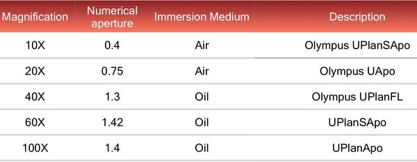
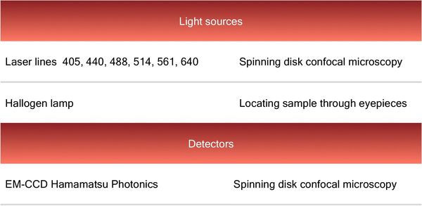
The UltraView system is mounted on an IX81 Olympus inverted microscope with environmental chamber for temperature and CO2 control. It is equipped with an electron-multiplying charge-coupled device camera (EM-CCD Hamamatsu Photonics) and it has 6 laser lines,  405, 440, 488, 514, 561 and 640 nm. This allows a very wide range of fluorescent probes to be used on the system. The system is run by Volocity software.

The instrument is also fitted with a photo-bleach/activation unit, which can be used to provide either single spots or arbitrary shapes via line or raster scans, suitable for FRAP, photodamage and photoactivation experiments.  The automated XYZ stage can be programmed for image stitching of large samples and time-lapse imaging at multiple positions.


For more information regarding training and booking please contact micron@bioch.ox.ac.uk.


Applications

  • Live cell imaging (temperature and CO2 control)
  • Optical sectioning for 3D imaging
  • Multi-channel XYZ fluorescence and transmitted light imaging modalities
  • Time-lapse imaging
  • Tile scanning and stiching 
  • Multipoint visiting with multiwells overlay
  • FRAP, Photoactivations and colocalisation

Technical specifications

Objectives

Light sources and Detector

Light sources and Detector

image374

Light sources and Detector

Light sources and Detector

Light sources and Detector

image375

Additional Information

Micron_PE_Spinning Disk_Manual (pdf)

Download

Micron access to Volocity (pdf)

Download

Copyright © 2018 Micron Oxford - All Rights Reserved.